Congenital Heart Diseases - 1
Congenital heart diseases are among the most common of all congenital malformations. In routine clinical practice, paediatricians and healthcare professionals encounter a wide variety of presentations, such as:
A term baby who had a normal new-born check, was discharged home, and later presents to the emergency department in shock
A term baby on the postnatal ward who is noticed to be cyanosed, yet otherwise appears well
A few-week-old infant presenting with recurrent chest infections, poor weight gain, feeding difficulties, and breathing problems
A referral from primary care due to the detection of a heart murmur in an otherwise stable baby.
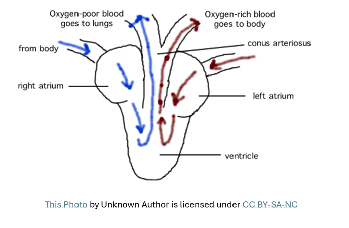
Normal Heart - Let me be a bit stupid and have a look at the very basics of circulation. Simplest of anatomy and physiology helps us in many ways, especially cardiac conditions.
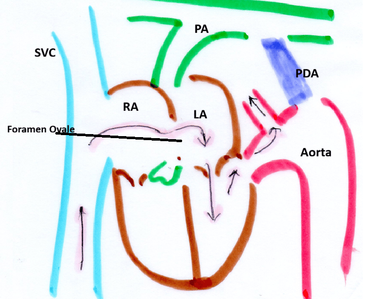
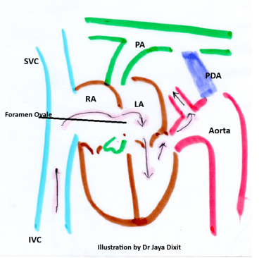
1. Two big veins SVC and IVC bring deoxygenated blood from the body to the Right Atrium.
Through Tricuspid Valve blood goes into the right ventricle.
The right ventricle pumps blood into pulmonary arteries via the pulmonary valve. (Ventricular systole)
Oxygenated blood from Lungs is then brought to the Left Atrium via Pulmonary veins.
From Left Atrium this blood goes in to Left Ventricle via Mitral Valve
The left ventricle pumps this blood into Systemic circulation via Aortic valve.
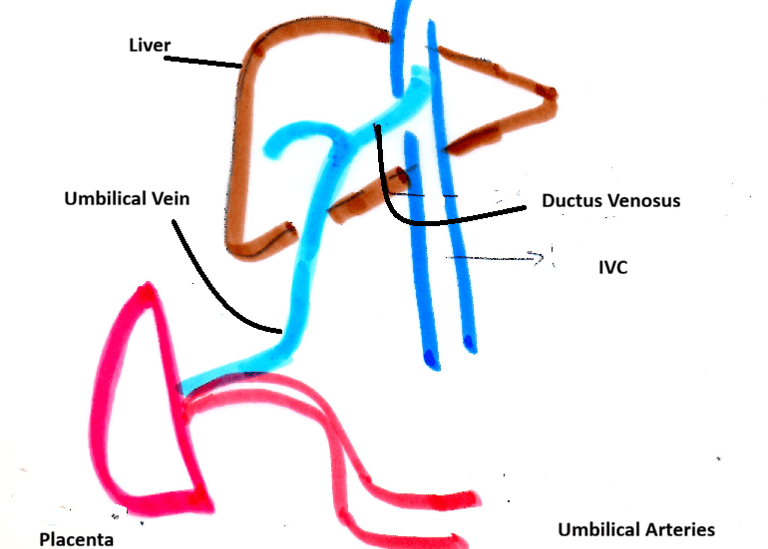
Foetal Circulation- Some understanding of foetal circulation is really important to understand the basic pathophysiology of some of the congenital cardiac lesions. Placenta is an an Organ, functioning as lungs, kidneys,, and gut. Also producing essential hormones. As we all know baby is connected by Umbilical Cord, it has two umbilical arteries and one umbilical vein
It is probably better to divide the Umbilical Circulation in 3 parts as below.
The umbilical vein carries relatively oxygenated blood from the placenta to the baby. About 50% of this blood passes through the liver, while the remaining 50% enters the inferior vena cava.
Inferior Vena Cava and Foramen Ovale
Blood from the inferior vena cava enters the right atrium but preferentially flows through the foramen ovale into the left atrium. From there, it enters the left ventricle and is pumped into the systemic circulation, mainly supplying the upper part of the body, including the brain.
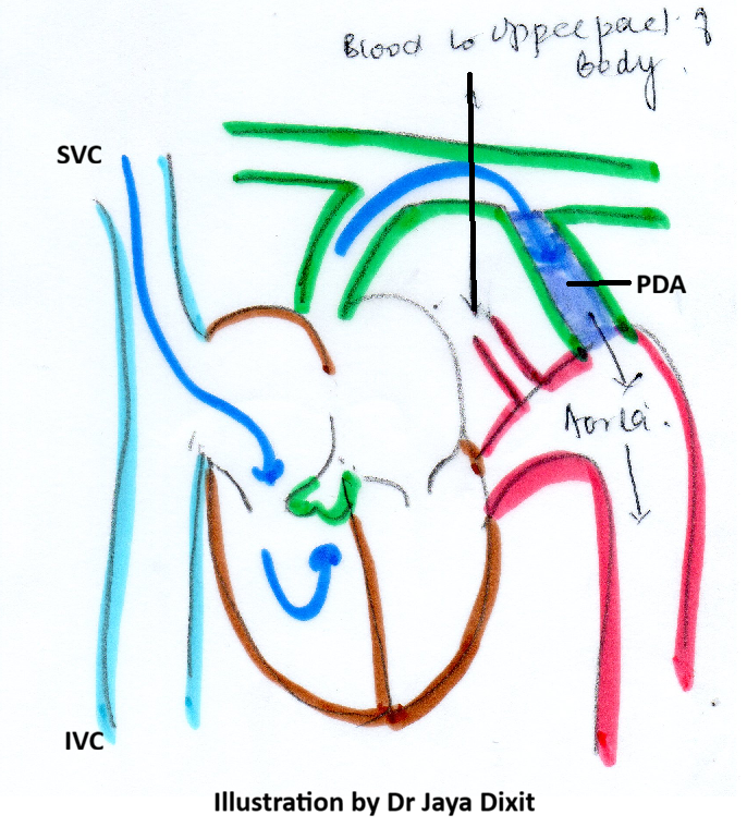
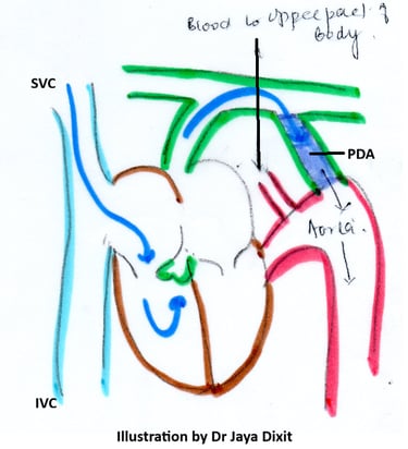
Superior Vena Cava and Ductus Arteriosus
Blood returning via the superior vena cava enters the right atrium and preferentially flows into the right ventricle, then into the pulmonary artery.
Because the foetal lungs are not functioning, only about 10% of this blood goes to the lungs. The majority bypasses the lungs through the patent ductus arteriosus (PDA) and enters the systemic circulation, supplying the lower part of the body.
Transition at Birth:
At birth, the baby is separated from the placenta when the umbilical cord is clamped. With the first breath:
Pulmonary vascular resistance drops significantly
Pulmonary blood flow increases dramatically
The lungs begin oxygenating blood
As a result:
Oxygenated blood returns to the left atrium and left ventricle
The foramen ovale closes due to pressure changes
The patent ductus arteriosus and ductus venosus constricts
The foetal circulation gradually transitions to normal postnatal circulation.
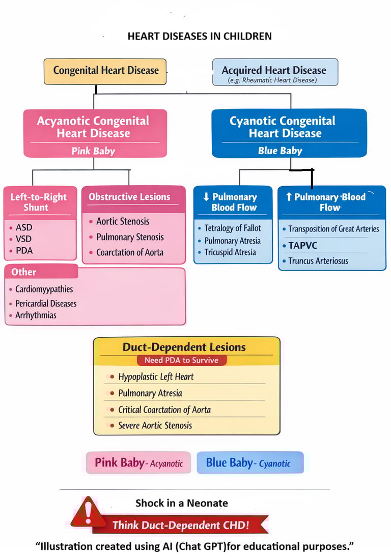
Classification of Heart Diseases in Children.
One of simplest way of to approach towards heart diseases in children is classify them. It helps in understanding their basic pathophysiology and also helps us to remember them better.